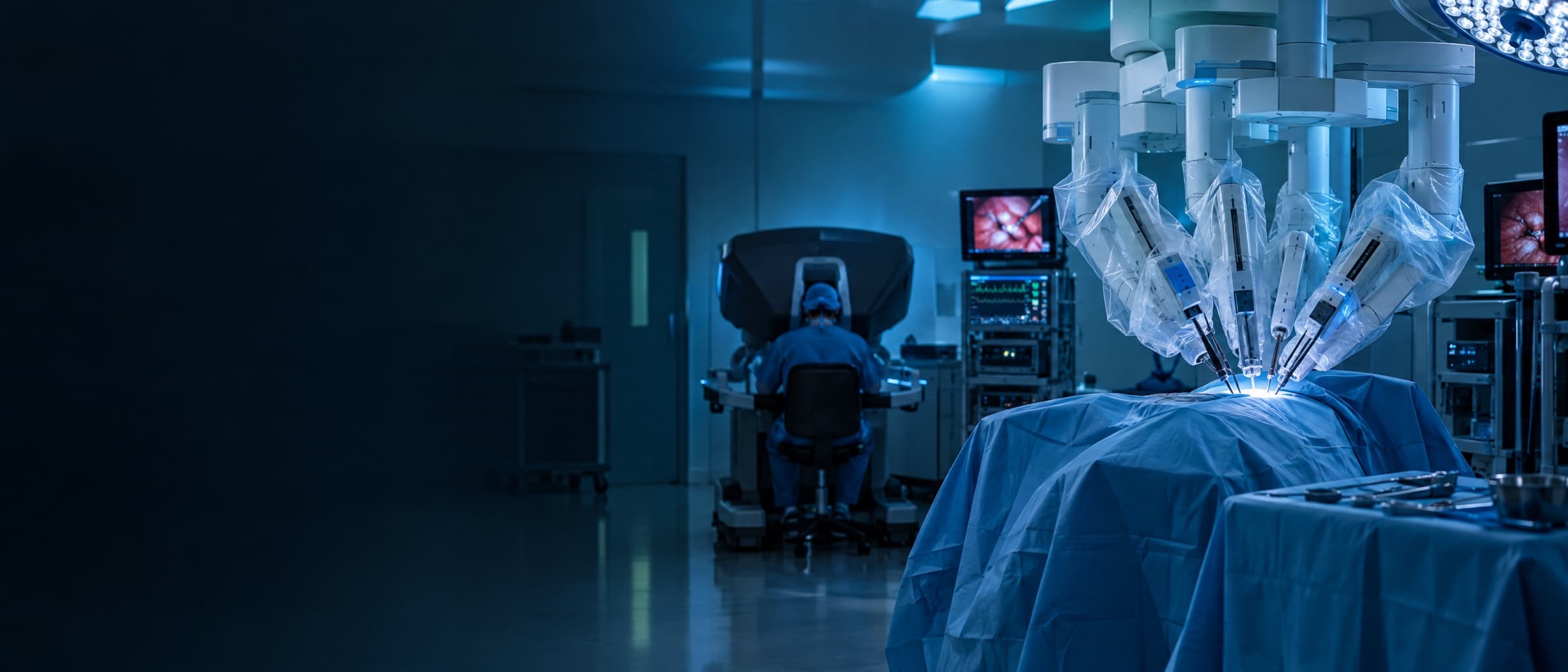

Real-time anatomy recognition in the robotic OR, built on surgeon-reviewed labels.
Read the story- annotated surgical frames
- 1,500+
- prototype on the robot console
- Live
Beyond Vision: CVAT Goes Multimodal and Introduces Audio Annotation. Learn more

CVAT turns surgical video, medical scans, and microscopy into validated training data for detection, segmentation, and clinical decision support, trusted by teams like UIC Urology.
Use cases
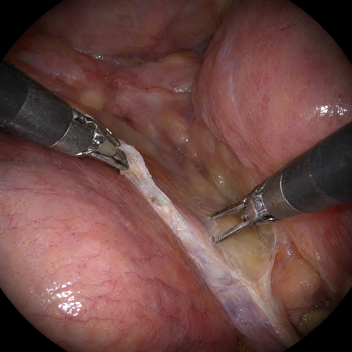
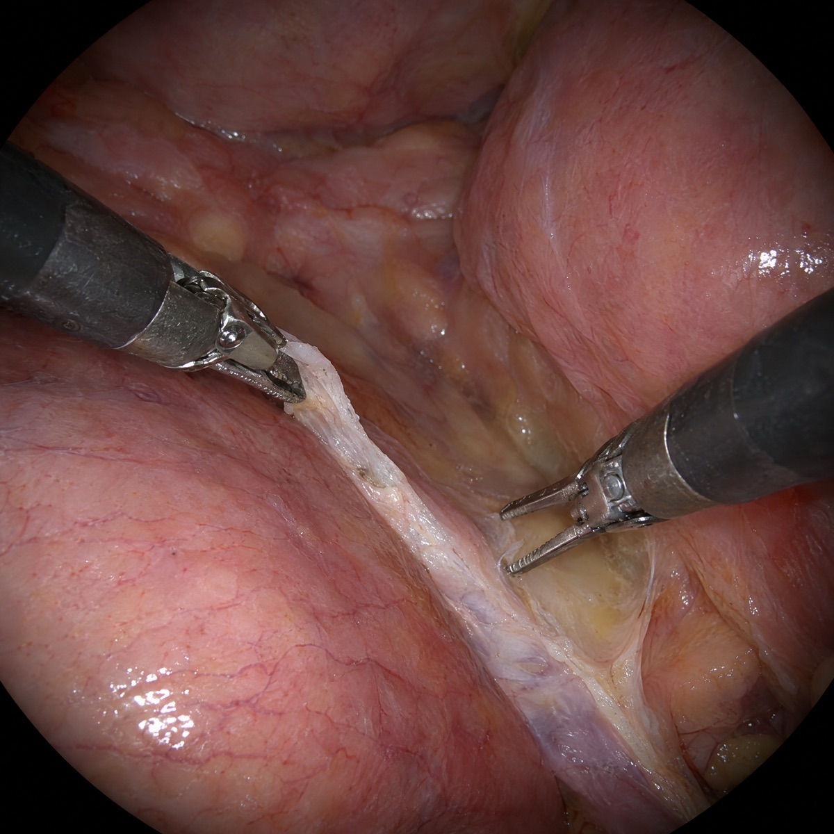
Segment anatomy and instruments frame by frame in surgical video.
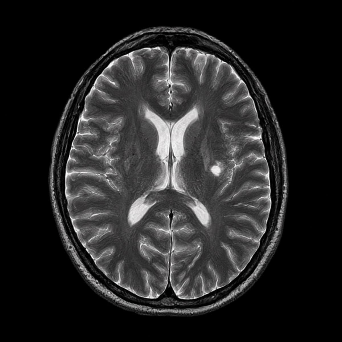
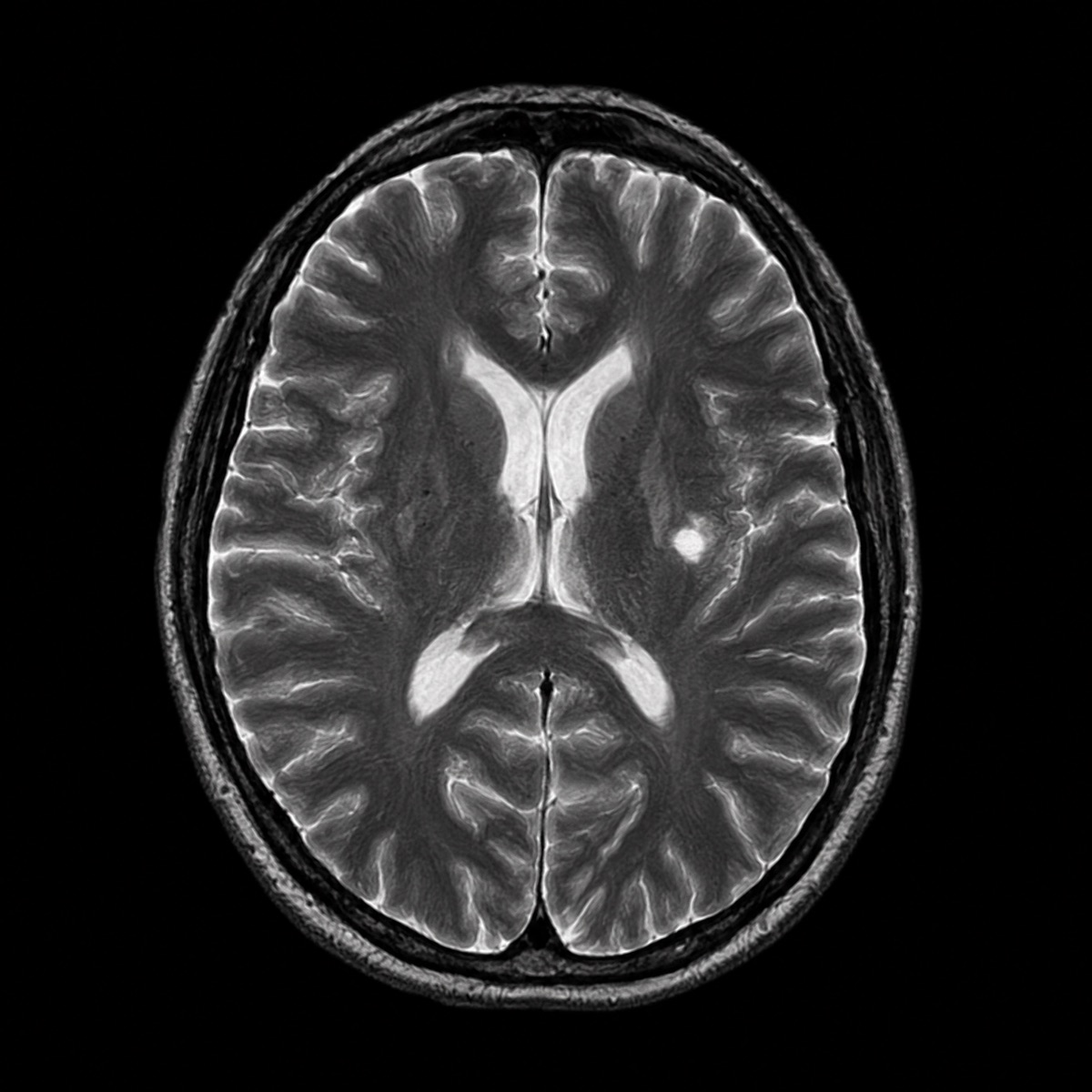
Outline organs, lesions, and nodules across X-ray, CT, and MRI.
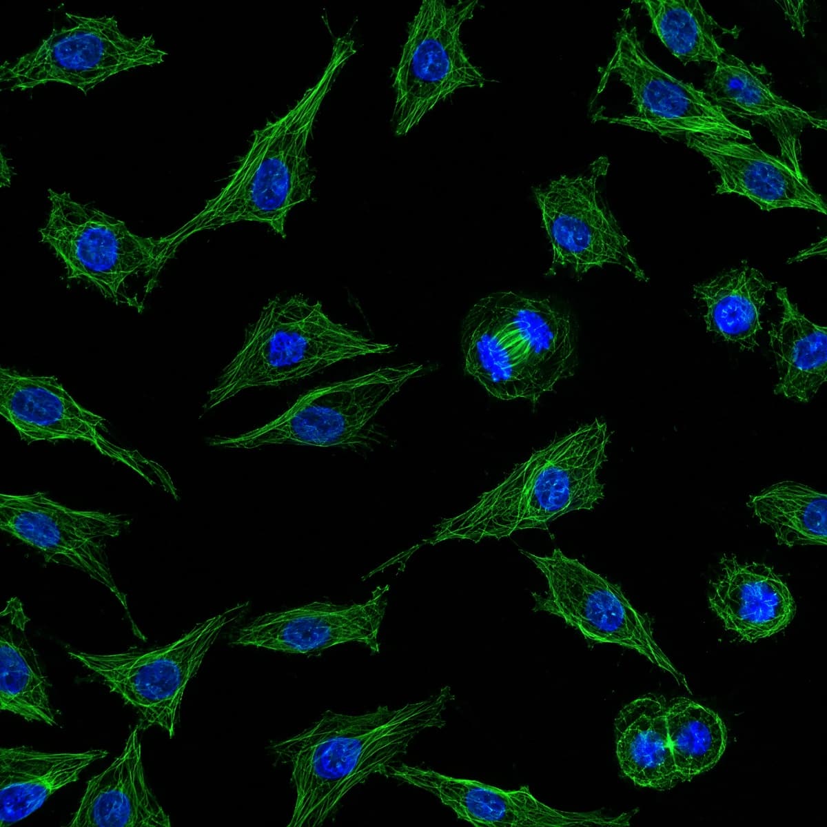
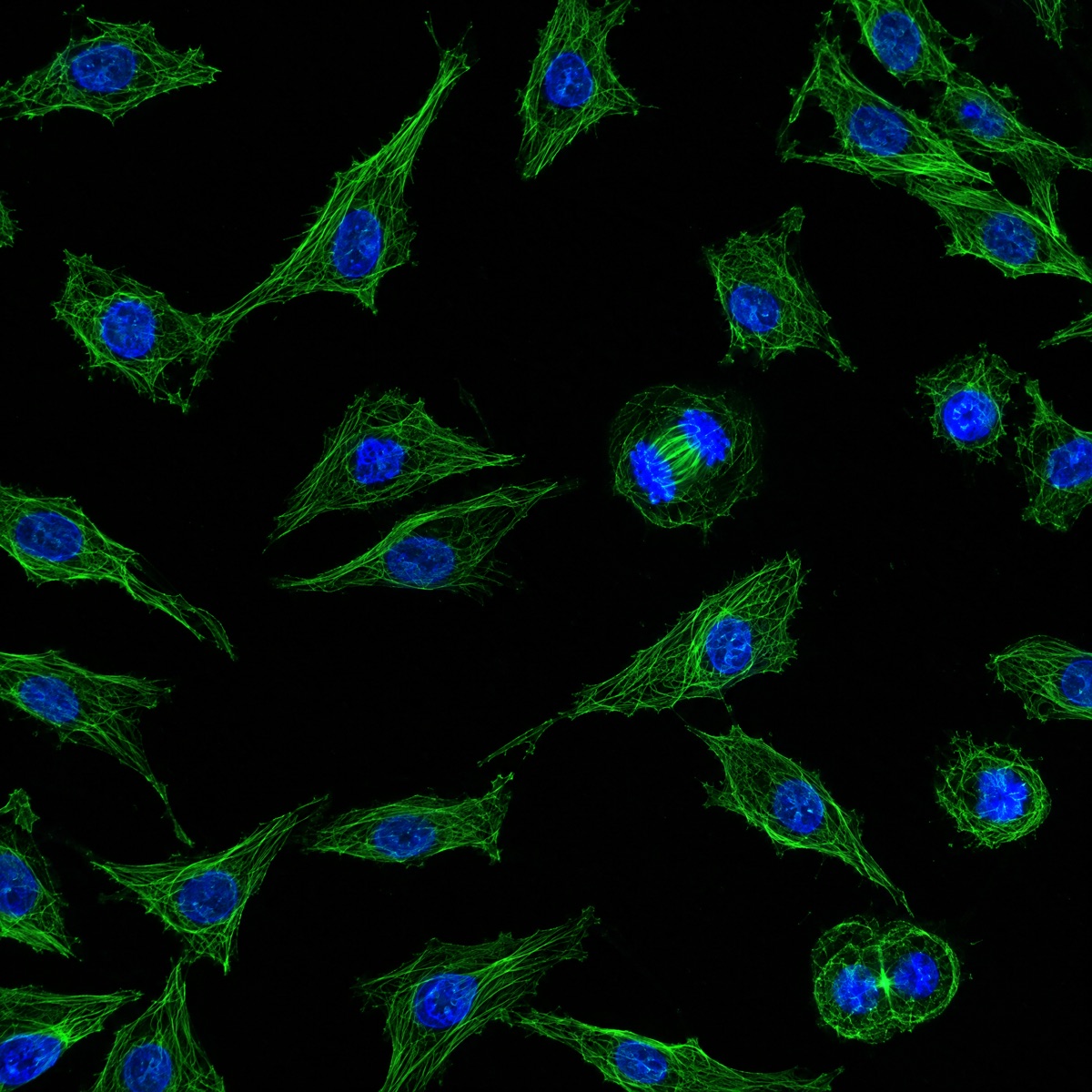
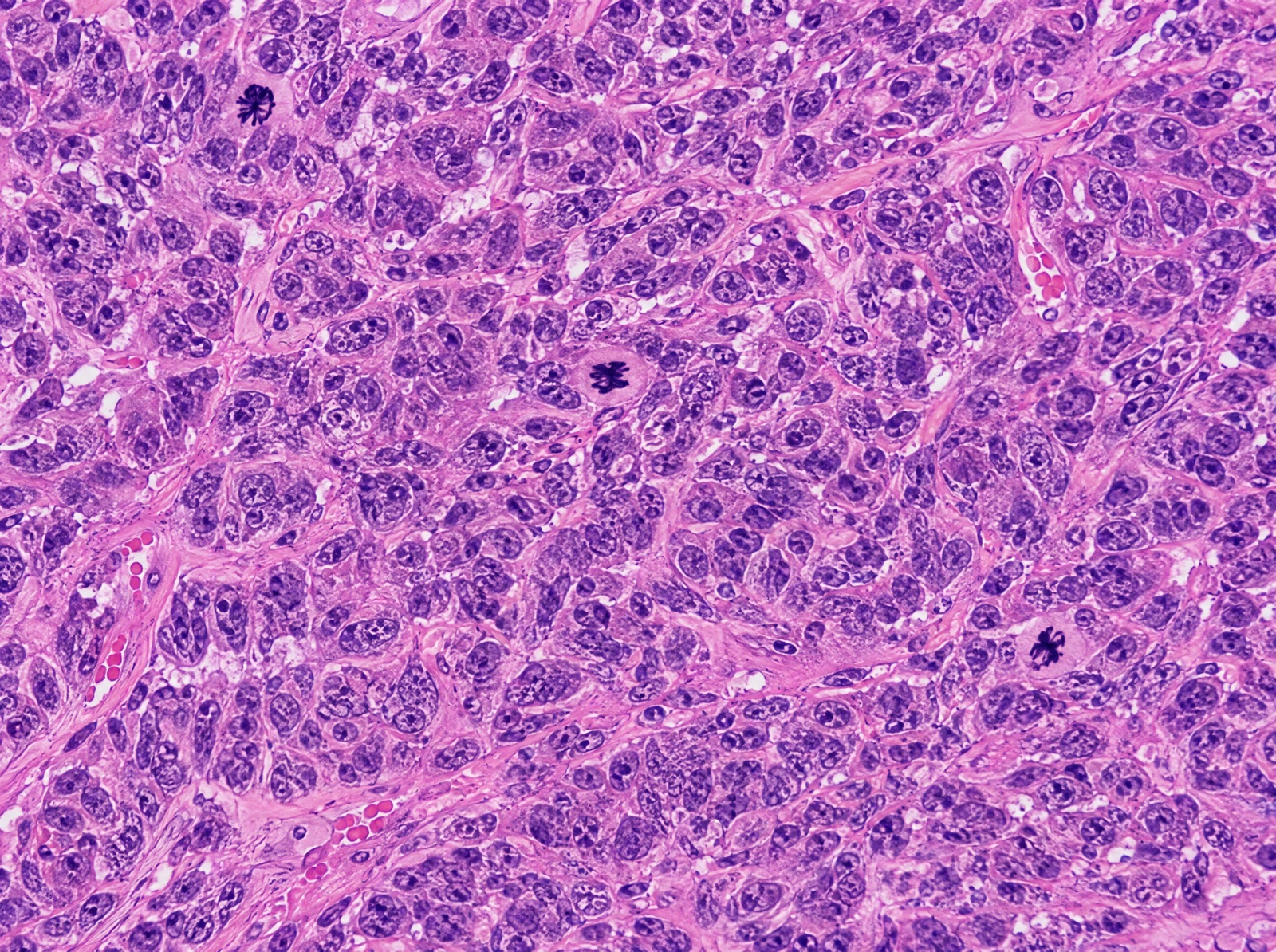
Count cells, nuclei, and dividing cells across every slide and well.
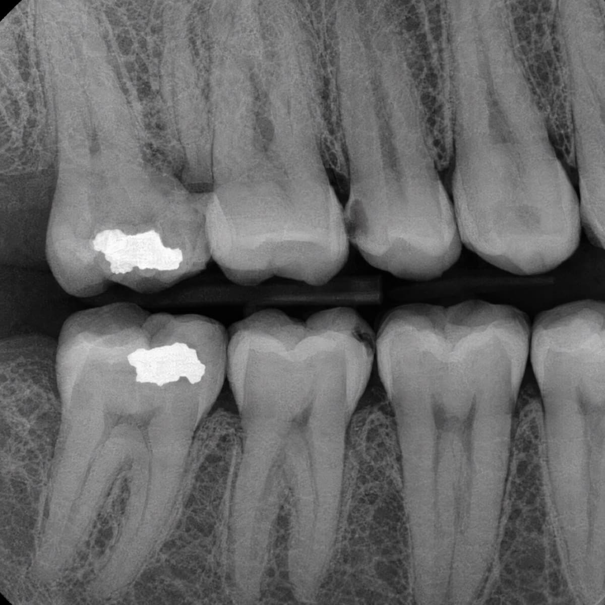
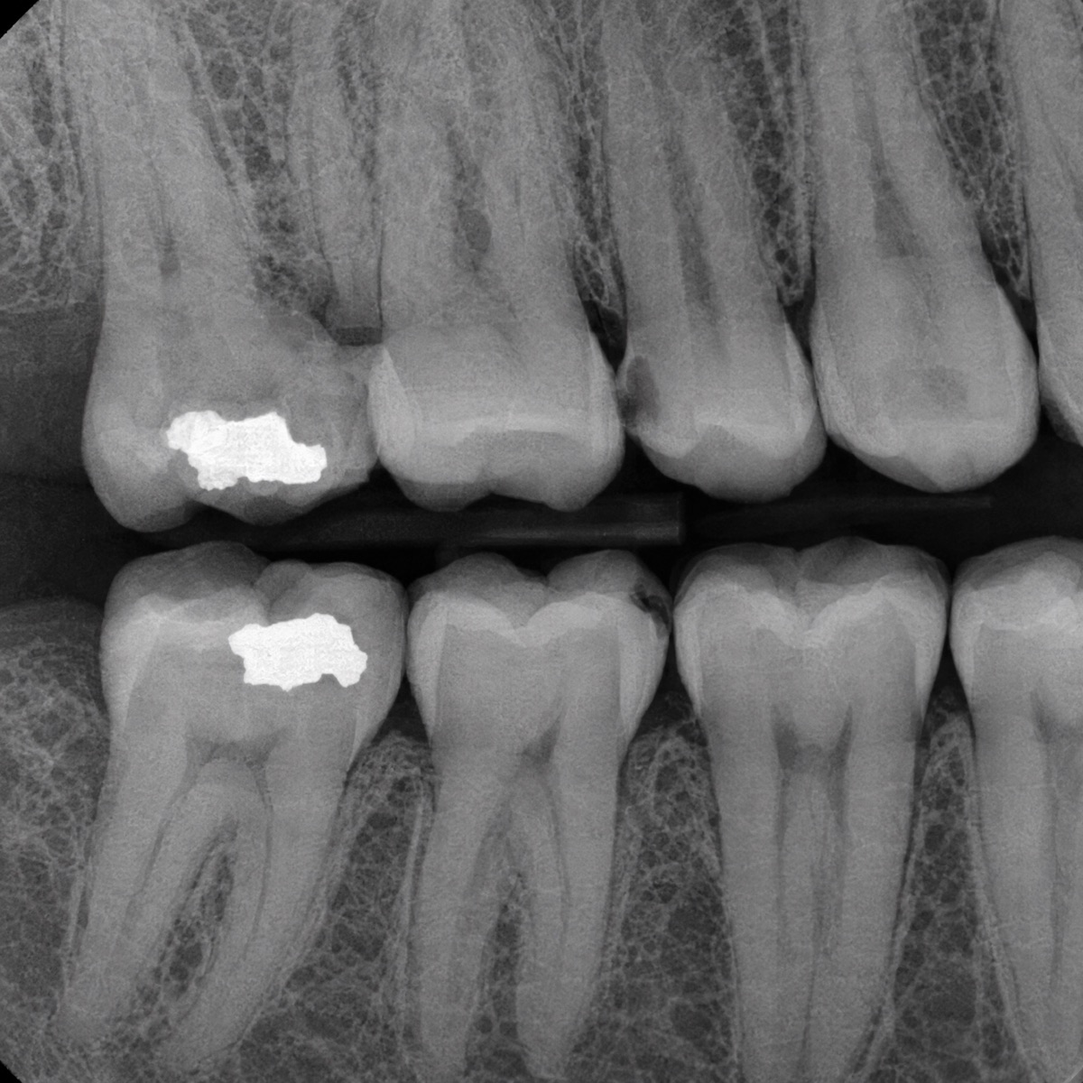
Label teeth, restorations, and caries on dental X-rays.
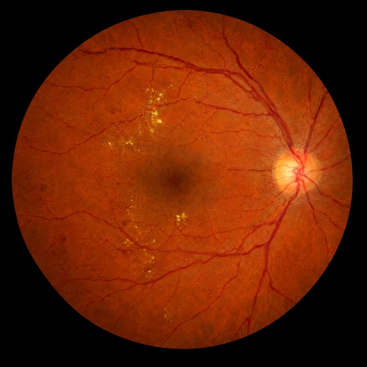
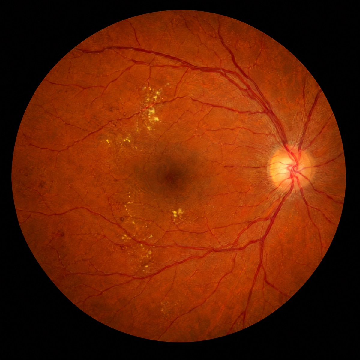
Mark the optic disc, vessels, and lesions in fundus photos.
Integrate Seamlessly
Connect your de-identified imaging archive, existing labels, and review stages as they are. Nothing to re-plumb.
Book a demoAccelerate Validation
AI pre-labels every frame and scan. Your clinicians review and fix what it misses.
Book a demo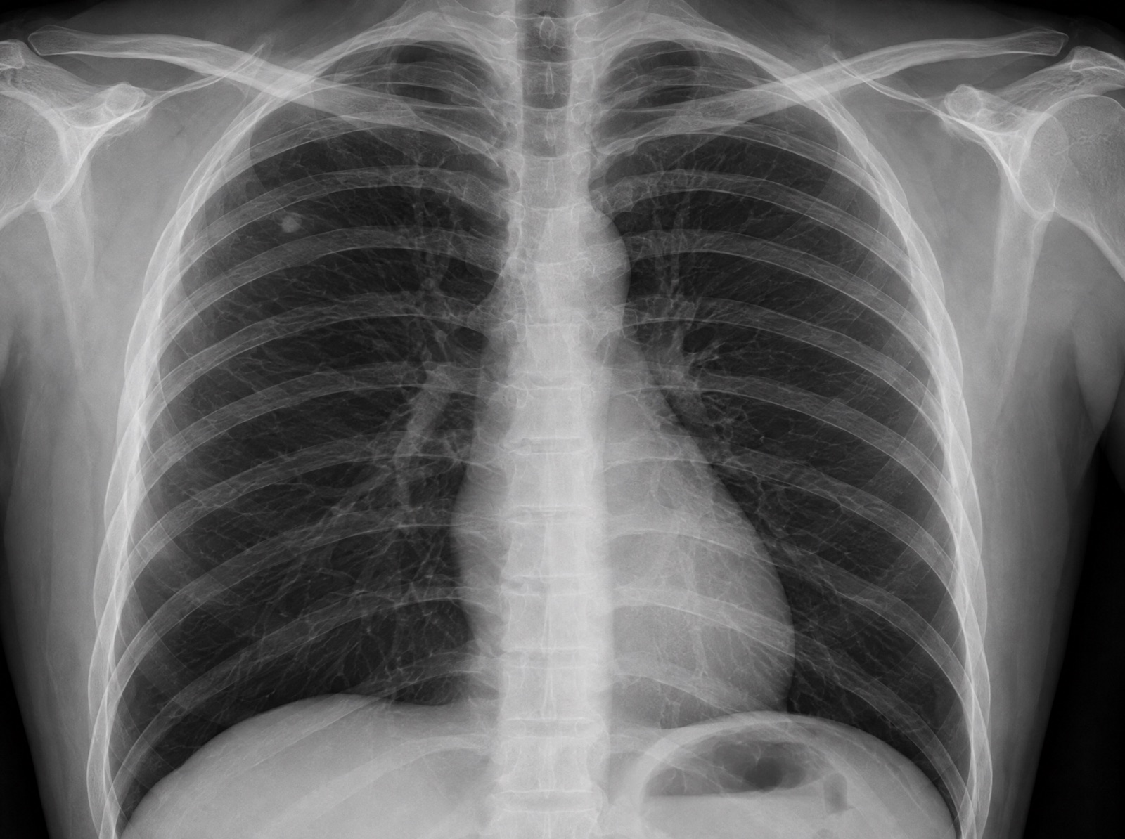
Ship with Confidence
Every job is scored against ground truth, so only clean data reaches training.
Book a demoMean annotation quality
98.4%
Target 95%
Honeypots
Real-time anatomy recognition in the robotic OR, built on surgeon-reviewed labels.
Read the story